Arachnoid Cyst
Arachnoid Cyst
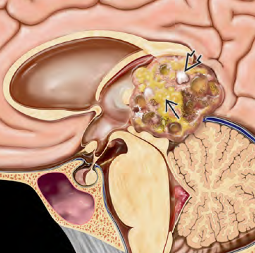
A pineal arachnoid cyst is a fluid-filled, benign lesion that arises from the arachnoid membrane in the region of the pineal gland, located near the center of the brain, adjacent to the third ventricle and the cerebral aqueduct. Though often asymptomatic and incidental, these cysts can sometimes grow in size and exert pressure on surrounding brain structures, leading to hydrocephalus and other neurological symptoms, especially in children. Arachnoid cysts in the pineal region are rare, but due to their proximity to vital neural pathways and CSF channels, early detection and neurosurgical assessment are crucial. In pediatric cases, timely intervention may prevent long-term neurological complications and improve quality of life. 🌍 Pineal Arachnoid Cysts in Bangladesh: The Need for Awareness In Bangladesh, pineal arachnoid cysts are often underdetected due to limited access to MRI imaging in many districts and upazilas. Children presenting with morning headaches, visual disturbances, or nausea are frequently treated symptomatically for gastritis, eye strain, or general weakness, without identifying the underlying neurological issue. Dr. Md. Nafaur Rahman, a leading pediatric neurosurgeon in Bangladesh, has seen numerous such cases where a simple imaging and timely intervention could have prevented vision loss, hydrocephalus, or school performance decline. At NINS and the Bangladesh Paediatric Neurocare Centre, such cases are evaluated using advanced diagnostic tools and managed through minimally invasive techniques when needed. 🧬 What Causes a Pineal Arachnoid Cyst? Congenital malformation of the arachnoid membrane May result from abnormal CSF accumulation between arachnoid layers Not considered a tumor, and non-cancerous Rarely acquired after trauma, infection, or hemorrhage 🧒 Common Symptoms in Children Most small cysts are asymptomatic, but larger cysts or those causing ventricular compression can present with: Morning headache (due to raised intracranial pressure) Nausea and vomiting Blurred or double vision, sometimes with upward gaze palsy (Parinaud’s syndrome) Lethargy or irritability Unsteady gait or poor balance Learning difficulties or behavior changes In infants, increasing head size or bulging fontanelle (soft spot) In Bangladesh, these symptoms may go unrecognized or misattributed, causing critical delays in neurosurgical referral. 🔍 Diagnosis of Pineal Arachnoid Cyst 🧠 MRI Brain (Preferred) Shows a well-demarcated, fluid-filled lesion in pineal region Hypointense on T1, hyperintense on T2, no enhancement post-contrast No solid components (helps distinguish from tumors) Often associated with dilated ventricles (hydrocephalus) 🧪 Differential Diagnosis Pineal cyst vs. pineal tumor (e.g., pineocytoma, germinoma) Cystic tumors or colloid cysts Tumor markers (AFP, β-HCG) in blood/CSF may be tested to rule out neoplasms 🛠️ Treatment Approach 1. Observation If cyst is small and asymptomatic, regular MRI monitoring is advised Clinical follow-up every 6–12 months 2. Surgical Intervention Indicated if the cyst causes: Hydrocephalus Parinaud’s syndrome Persistent headache or visual issues 🧪 Surgical Options: Endoscopic Third Ventriculostomy (ETV): Preferred for hydrocephalus without touching the cyst Minimally invasive, avoids permanent shunt Endoscopic Fenestration of Cyst: Creates opening in cyst wall to allow drainage into CSF pathways Often combined with ETV VP Shunt: Used if ETV not feasible or failed Requires lifelong follow-up Dr. Md. Nafaur Rahman specializes in neuroendoscopic procedures for pineal region cysts, minimizing surgical risk and promoting faster recovery. 🔄 Prognosis Excellent prognosis after successful ETV or cyst fenestration Most children resume normal activities and schooling Rare recurrence if properly treated No chemotherapy or radiation required Lifelong monitoring may not be necessary if symptoms resolve and cyst remains stable ⚠️ Complications of Delayed Diagnosis Hydrocephalus and pressure effects on midbrain Vision problems or permanent gaze palsy Seizures (rare) Developmental delay, poor school performance Psychological stress for family and child 👨⚕️ Why Choose Dr. Md. Nafaur Rahman? One of Bangladesh’s leading pediatric neurosurgeons Expertise in endoscopic neurosurgery, pineal region surgery, and CSF diversion techniques Provides multidisciplinary care with neurologists, radiologists, and pediatricians Based at National Institute of Neurosciences & Hospital (NINS) Offers specialized services through Bangladesh Paediatric Neurocare Centre 📞 Book an Appointment Dr. Md. Nafaur Rahman Assistant Professor, Pediatric Neurosurgery, National Institute of Neurosciences & Hospital (NINS) Chief Consultant, Bangladesh Paediatric Neurocare Centre 📞 Serial & Appointment: 01912988182 | 01607033535 🌐 Website: www.neurosurgeonnafaur.com


