Tectal Plate Glioma
Tectal Plate Glioma
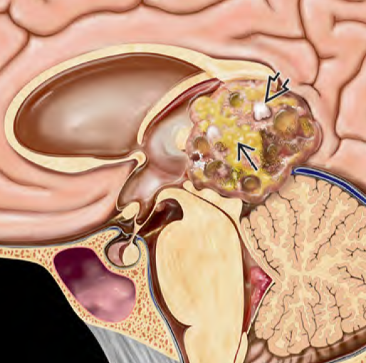
Tectal Plate Glioma, also known as tectal glioma or tectal gliosis, is a slow-growing, low-grade brain tumor located in the tectum (roof) of the midbrain, near the aqueduct of Sylvius. Although rare, it predominantly affects children and adolescents, and unlike other brainstem gliomas, it typically exhibits an indolent course with minimal symptoms apart from hydrocephalus. These tumors are usually WHO Grade I pilocytic astrocytomas and are often discovered incidentally during evaluation of increased intracranial pressure. In most cases, surgical removal is not required unless there is progression. Instead, long-term surveillance with MRI and management of hydrocephalus are the primary approaches. 🌍 Bangladesh Perspective on Tectal Plate Glioma In Bangladesh, many children with tectal gliomas remain undiagnosed or misdiagnosed due to lack of access to advanced neuroimaging like MRI brain with contrast. Symptoms such as morning headache, vomiting, and blurred vision are often misattributed to migraine, vision problems, or GI disorders. As a result, delays in appropriate neurosurgical referral are common. At the National Institute of Neurosciences & Hospital (NINS) and Bangladesh Paediatric Neurocare Centre, Dr. Md. Nafaur Rahman offers specialized diagnostic, surgical, and follow-up services for children with tectal gliomas and other brainstem tumors. 🧬 Pathophysiology Originates from astrocytes in the tectum of the midbrain Causes compression of the cerebral aqueduct, leading to obstructive hydrocephalus Tumors are typically non-enhancing, small, and stable in size Histologically classified as pilocytic astrocytoma or low-grade glioma (WHO Grade I or II) Rarely undergoes malignant transformation or spreads 🧒 Symptoms of Tectal Plate Glioma Most symptoms are related to hydrocephalus, caused by obstruction of cerebrospinal fluid (CSF) flow through the aqueduct. Common signs include: Morning headache Nausea and vomiting Blurred or double vision Papilledema (optic disc swelling) Parinaud's syndrome: inability to look upwards, pupillary disturbances Unsteady gait, fatigue Changes in behavior or school performance In Bangladesh, these symptoms may go unrecognized or be managed symptomatically for months before imaging is advised. 🔍 Diagnostic Approach 🧠 MRI Brain with Contrast Key investigation Reveals a small lesion in the tectal plate with or without mild contrast enhancement Shows dilated lateral and third ventricles, confirming hydrocephalus Spinal MRI may be done to rule out drop metastasis in rare cases 👁️ Fundoscopy May reveal papilledema in children with raised intracranial pressure 🚫 Biopsy Not usually performed unless there is progression or diagnostic uncertainty due to the risk of damage to brainstem structures 🛠️ Treatment Options 💧 Management of Hydrocephalus Endoscopic Third Ventriculostomy (ETV): preferred method Creates a bypass for CSF flow and eliminates need for long-term shunt Dr. Nafaur Rahman has expertise in performing ETV using modern endoscopic systems Ventriculo-Peritoneal (VP) Shunt: alternative for cases where ETV is not feasible Requires long-term follow-up for shunt function and complications 🩺 Observation Strategy Tumor itself is usually not operated unless symptomatic or growing MRI surveillance every 6 months to 1 year Clinical monitoring of vision, gait, and school performance 🧪 Surgery or Biopsy (Rare) Indicated only in atypical or progressive lesions May require stereotactic or open biopsy 🔄 Prognosis Excellent long-term outlook Many children lead normal lives with stable tumor after CSF diversion Low risk of progression or malignant transformation Need for surgery on the tumor itself is rare Regular follow-up is essential to monitor for signs of progression ⚠️ Risks of Delayed Diagnosis Raised intracranial pressure leading to optic nerve damage and vision loss Chronic hydrocephalus causing cognitive delay Missed opportunity for simple ETV procedure, leading to emergency situations Long-term neurological damage due to brainstem compression 👨⚕️ Why Choose Dr. Md. Nafaur Rahman? Recognized expert in pediatric brainstem and pineal region tumors in Bangladesh Advanced training in neuroendoscopy, ETV, and minimally invasive neurosurgery Accurate diagnosis using state-of-the-art imaging and monitoring tools Individualized, child-focused care plans and long-term follow-up Based at National Institute of Neurosciences & Hospital (NINS) and Bangladesh Paediatric Neurocare Centre 📞 Contact for Appointment Dr. Md. Nafaur Rahman Assistant Professor, Pediatric Neurosurgery, National Institute of Neurosciences & Hospital (NINS) Chief Consultant, Bangladesh Paediatric Neurocare Centre 📞 Serial & Appointment: 01912988182 | 01607033535 🌐 Website: www.neurosurgeonnafaur.com