Teratomas
Teratomas
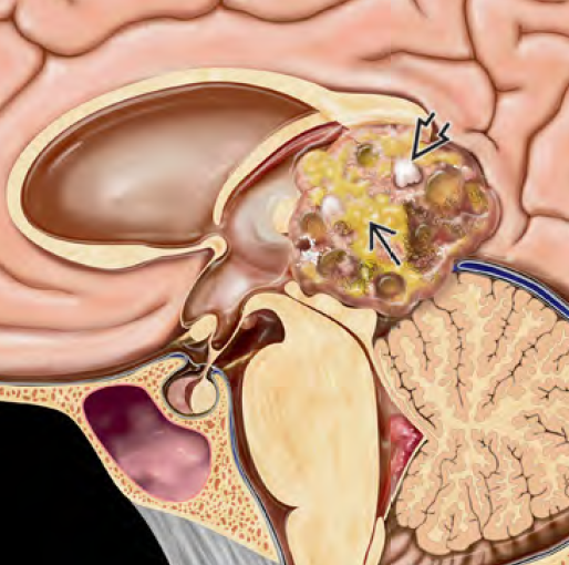
A teratoma is a type of germ cell tumor that contains tissues from all three embryonic layers: ectoderm, mesoderm, and endoderm. These tumors can be mature (benign) or immature/malignant, depending on their histological appearance. In children, teratomas may appear in the brain (intracranial teratoma) or along the spinal cord (intraspinal teratoma) and are congenital in many cases, forming during fetal development. Pediatric teratomas are rare but serious, particularly if they occur in vital CNS areas, as they may lead to neurological deficits, hydrocephalus, seizures, or even life-threatening complications. Early diagnosis, timely surgical intervention, and long-term monitoring are essential to ensure the best outcome. 🌍 Teratomas in the Bangladesh Perspective In Bangladesh, pediatric teratomas are underdiagnosed due to limited prenatal imaging, lack of pediatric neuro-oncology awareness, and delayed referral to specialized neurosurgical centers. Many children born with cranial or spinal masses are first seen by general physicians or local hospitals, where such conditions may be misinterpreted as infections, birth trauma, or simple cysts. Dr. Md. Nafaur Rahman, one of the leading pediatric neurosurgeons in Bangladesh, offers comprehensive diagnosis and surgical care for infants and children with brain and spinal teratomas at the National Institute of Neurosciences & Hospital (NINS) and the Bangladesh Paediatric Neurocare Centre. 🧬 Types of Teratomas in Children Mature Teratoma Benign Contains differentiated tissues like hair, teeth, fat, or cartilage May still cause compression symptoms in the brain or spine Immature Teratoma Malignant potential Contains embryonic tissue components Requires surgery plus chemotherapy Mixed Germ Cell Tumor with Teratomatous Component Contains teratoma with other germ cell elements (e.g., yolk sac, choriocarcinoma) 🧠 Common Locations Intracranial Teratomas Pineal region Suprasellar/hypothalamic area Cerebral hemispheres Posterior fossa Can present antenatally or neonatally Spinal Teratomas Intradural or extradural Cervical, thoracic, or lumbosacral spine Often associated with spinal dysraphism (e.g., spina bifida, dermal sinus) 🧒 Symptoms of Pediatric Teratomas Intracranial: Large head or bulging fontanelle in neonates Hydrocephalus and vomiting Seizures Weakness or poor feeding Developmental delay Cranial nerve palsy Endocrine disturbances (if in suprasellar region) Spinal: Swelling or mass over spine Weakness in legs Bowel or bladder dysfunction Tethered cord syndrome Association with skin tags, hairy patches, or dimples In Bangladesh, such signs are often overlooked or misdiagnosed as hydrocephalus, cystic swelling, or benign birthmarks, especially in rural areas. 🔍 Diagnostic Approach 🧠 Imaging: MRI of brain/spine with contrast – gold standard Shows complex mixed-density lesion with cystic, fatty, and sometimes calcified components Teratomas often cause mass effect, hydrocephalus, or spinal cord compression 💉 Tumor Markers: AFP (Alpha-fetoprotein) and β-hCG may be elevated in malignant forms Helps differentiate from other germ cell tumors 🧬 Histopathology: Confirms diagnosis and determines grade (mature vs immature) Required for treatment planning 🛠️ Treatment Options ✂️ Surgical Resection: Mainstay of treatment Goal: Maximal safe removal of tumor Performed using microsurgical techniques, neuro-navigation, and intraoperative monitoring Dr. Nafaur Rahman has significant experience in removing both cranial and spinal teratomas in neonates and young children 💊 Chemotherapy: Reserved for immature or malignant teratomas Protocols based on international pediatric oncology guidelines ☢️ Radiotherapy: Rarely needed in young children unless tumor recurs or cannot be completely resected 🔄 Prognosis Mature teratomas – excellent prognosis with complete removal Immature/malignant teratomas – prognosis depends on tumor location, size, and histology Long-term follow-up needed for recurrence monitoring, especially in spinal teratomas with tethered cord risk ⚠️ Delayed Treatment Risks Increased intracranial pressure leading to herniation Neurological damage due to spinal cord compression Permanent visual or hormonal impairment Hydrocephalus requiring VP shunt Tumor rupture or infection Malignant transformation if untreated 👨⚕️ Why Dr. Md. Nafaur Rahman? National expert in pediatric congenital brain and spinal tumors Advanced neurosurgical training in teratoma resection Uses latest intraoperative technologies for safe tumor removal Offers full-spectrum care: diagnosis, surgery, follow-up, and rehabilitation Based at NINS, and available at Bangladesh Paediatric Neurocare Centre 📞 Book an Appointment Today Dr. Md. Nafaur Rahman Assistant Professor, Pediatric Neurosurgery, National Institute of Neurosciences & Hospital (NINS) Chief Consultant, Bangladesh Paediatric Neurocare Centre 📞 Serial/Appointment: 01912988182 | 01607033535 🌐 Website: www.neurosurgeonnafaur.com